A psychologist in Brazil spent weeks treating what she assumed was migraine with aura before a scan revealed a lesion in her cerebellum, UOL reports. The case, published by the outlet on October 28, 2026, illustrates why brain tumors are often found by accident rather than through a clear set of early symptoms.
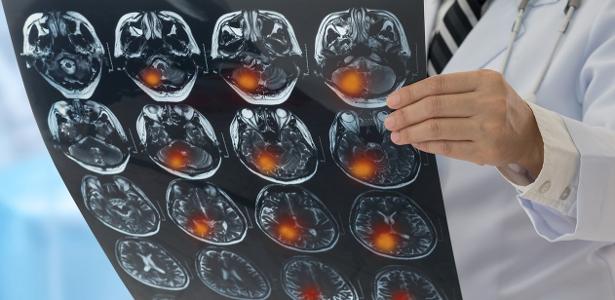
Nathalie De Pietro, 26, began having headache attacks roughly twice a week in early 2025, according to UOL. The episodes came with distorted images and blurred vision, and they interfered with her work during consultations with patients. She sought a neurologist, who ordered an MRI. The scan showed a lesion near the back of the head.
Further tests followed, and about three months after that first image she underwent surgery to remove a grade 1 pilocytic astrocytoma, a tumor classified as benign. UOL reports that the removal was complete, but her migraine attacks continued.
Why it matters
That detail carries a broader lesson, according to the specialists quoted by UOL: finding a lesion while investigating a symptom does not prove the lesion caused the symptom. Primary headaches such as migraine are common, and they frequently lead to scans that turn up tumors incidentally.
"Most frequently, a tumor is diagnosed as an imaging finding during the investigation of headache," Vinicius Trindade Gomes da Silva, the neurosurgeon who operated on Nathalie and leads the central nervous system tumor reference center at A.C. Camargo Cancer Center, told UOL.
Because brain tumors generally grow slowly, symptoms can emerge gradually and become obvious only once a lesion has reached a larger size, UOL reports.
Which headaches deserve a scan
Marcos Castello Barbosa de Oliveira, a neurologist at the Hospital das Clínicas of the USP medical school, told UOL that headaches unlike a person's usual pattern deserve investigation, particularly when they intensify over time.
He listed additional red flags: pain that wakes a person at night, pain that worsens with coughing or lying down, and pain that does not respond to painkillers. A new headache in someone over 50 should also raise suspicion of something more serious, he said.
UOL reports that people with no history of epilepsy who experience a first seizure should also have brain imaging to look for possible causes, tumors among them.
Behavioral shifts can be harder to spot. According to UOL, a person may become more withdrawn or more irritable, miss appointments, make mistakes on routine tasks, or struggle increasingly at work. Castello noted that such changes tend to progress faster than those seen in neurodegenerative conditions such as Alzheimer's, unfolding over days or months rather than years.
Location shapes the symptoms
Where a tumor sits matters, because different brain regions control different functions, UOL reports. A lesion in the left temporal region can affect speech and language, while one in the frontal region may alter behavior. A lesion in the occipital lobe can compromise part of the visual field. When a tumor affects one side of the brain, the weakness or other symptoms typically appear on the opposite side of the body, according to Castello.
The first step in evaluation is a conversation with a doctor and a neurological exam, UOL reports. If warning signs are present, the specialist may order imaging. A head CT with contrast is a fast option, especially in urgent situations, but MRI gives a more detailed view and, under ideal conditions, is the main exam for assessing a lesion. Advanced MRI techniques can help form a diagnostic hypothesis and guide treatment. After her first MRI, Nathalie underwent proton spectroscopy, which analyzes the biochemical characteristics of tissue.
Biopsy is not mandatory in every case, according to UOL. The neurosurgeon at A.C. Camargo said it is reserved mainly for situations where the diagnosis is uncertain or where the lesion cannot be removed safely without risking neurological harm.
Primary tumors versus metastases
UOL also draws a distinction between primary brain tumors and metastases. For a cancer cell to reach the brain, it must cross the blood-brain barrier, a structure that shields the brain and restricts the passage of substances and cells carried in the blood. Trindade told UOL that some tumors, particularly lung, breast and melanoma, carry cellular features such as adhesion proteins, enzymes and inflammatory substances that make crossing that barrier easier.
The two categories also behave differently. Primary tumors tend to infiltrate surrounding brain tissue, while metastases usually form more clearly defined lesions. Because metastatic cells arrive through the bloodstream, several regions can be affected at once. UOL notes that tumors of the skin and skull can also invade brain tissue through local growth.
What happens next
Surgery is the initial treatment in most cases, apart from small metastases that may be addressed with radiosurgery or targeted therapies, according to UOL. Trindade said the aim is to remove as much of the tumor as possible, preferably completely, while preserving the patient's neurological and functional status. The outlet's report was cut off mid-sentence as the neurosurgeon began describing the tools used to achieve that precision, so the full account of surgical technique was not available.
Watch for fuller reporting from UOL on surgical approaches, and for wider discussion among clinicians about when headache and behavioral changes should prompt brain imaging.