Researchers at the Allen Institute have found that a small cluster of nerve cells in the brainstem does not send one uniform chemical signal across the brain, as scientists had long assumed. Instead, according to a study published in Nature and reported by Science Daily, the region communicates through several separate pathways aimed at specific destinations. The work used brain imaging, neural recordings, genetic analysis and behavioral tests in mice, and was published in late October 2026.
The cluster is the locus coeruleus, whose Latin name means "blue place." It holds only a small share of the brain's neurons but produces norepinephrine, a chemical messenger involved in attention, learning, heart rate, and the body's response to stress and excitement.
Why it matters
Norepinephrine is a central target for medicines used against depression, anxiety and ADHD. Existing drugs that act on it can influence several brain circuits at once, which may help in one area while causing unwanted effects in another. A clearer picture of how the locus coeruleus routes its signals could help researchers design treatments that act on specific circuits tied to specific symptoms.
The cells of the locus coeruleus are also among the first in the brain to break down in Alzheimer's disease, Science Daily reports, which gives the findings possible relevance for understanding that condition as well.
A postal network, not a foghorn
The old model treated the locus coeruleus like a loudspeaker, pushing essentially the same norepinephrine message everywhere. The new work contradicts that. By tracing how the cells connect, the researchers found that different groups of neurons reach different parts of the nervous system, which decides where norepinephrine is released.
Cells in the upper, or dorsal, part of the cluster connect mainly to the cerebral cortex and forebrain. Cells in the lower, or ventral, part send their signals chiefly to the brainstem and spinal cord. That split lets separate parts of the locus coeruleus affect separate brain functions.
Karel Svoboda, director of Neural Dynamics at the Allen Institute and a co-author of the study, described the system as closer to a targeted postal network than a foghorn.
Learning versus engagement
The two groups of cells also behaved differently during experiments. Dorsal neurons linked to the cortex were especially active while mice learned. Their activity rose when the animals made decisions and changed their choices after a poor result.
Ventral neurons followed a different pattern. Their activity climbed shortly before mice disregarded cues that a reward might be available, which the researchers say points to a role in deciding whether an animal responds to opportunities around it at all.
Svoboda said the work produced a clear map: dorsal cells projecting upward to the cortex take part in learning, while ventral cells projecting downward to the brainstem and spinal cord control whether animals engage with their surroundings. He added that the anatomical differences are matched by distinct patterns of gene expression, meaning the two groups differ not only in location and wiring but in which genes they switch on.
Very long cells with narrow targets
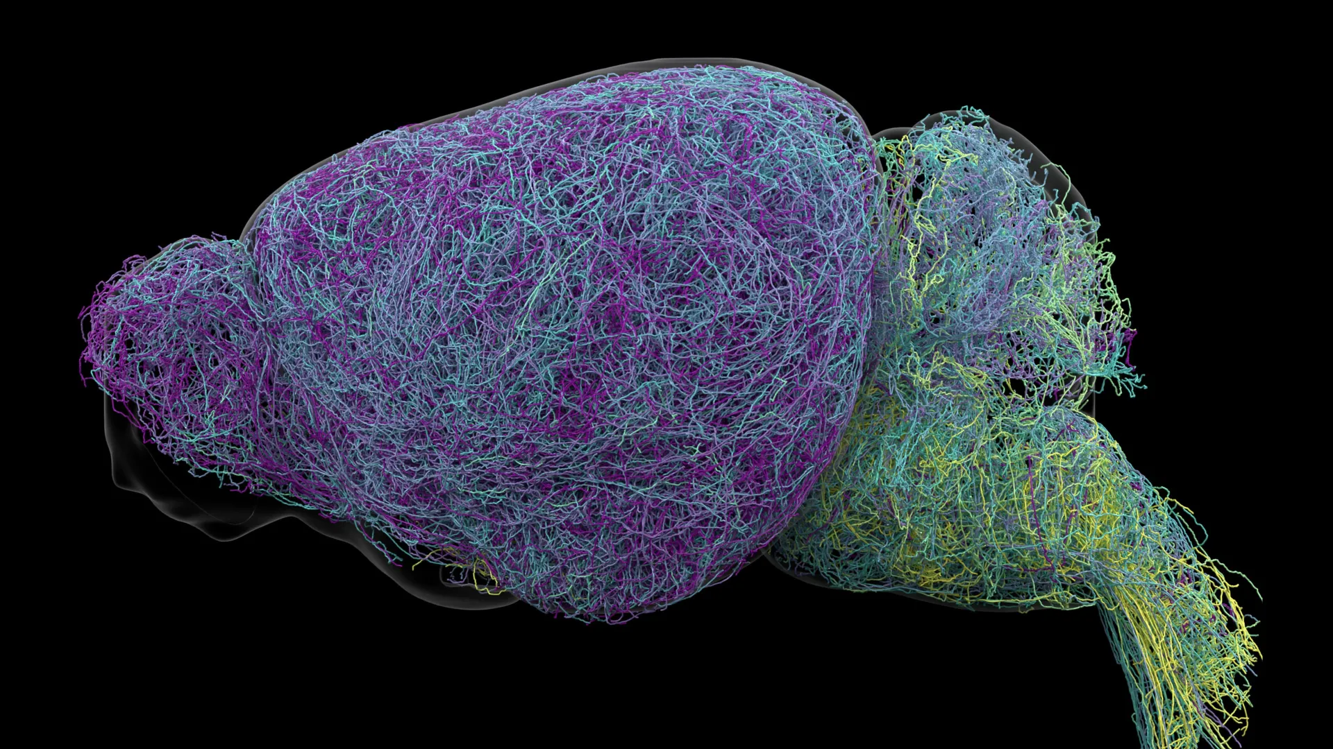
The study covered the mouse brain in detail. The team imaged nearly 35,000 neurons across the whole brain, examined the genetic profiles of almost 400,000 cells, and rebuilt the full shapes of several unusually complex nerve cells.
That reconstruction turned up axons, the projections that carry signals to other cells, averaging about 35 centimeters long. One axon measured 70.32 centimeters in total, placing it among the longest single neurons ever recorded in a mouse.
Jeremiah Cohen, another Allen Institute scientist and co-author, said this cell supplies norepinephrine to a very large volume of the cerebral cortex, which is unusual for the brain, since most neurons are more specific about their targets. Even so, he said, the neuron does not release norepinephrine everywhere: it skips the cerebellum, brainstem and spinal cord. Its reach is wide but still selective, which supports the finding that the locus coeruleus runs on carefully organized routes.
The researchers also noted parallels with the brain's dopamine system, which signals to the basal ganglia, a region involved in habit formation. Scientists suspect the two messengers cooperate in learning, possibly letting the brain pick up several complex or abstract ideas at once.
The study received support from the National Institutes of Health's Brain Research Through Advancing Innovative Neurotechnologies Initiative, known as The BRAIN Initiative.
What to watch next is whether these mouse findings hold in other species and whether drug developers can use the pathway map to build more selective treatments.